
Simple Staining- Principle, Procedure and Result Interpretation

Objectives of Simple Staining
- To perform a simple staining procedure.
- To compare the morphological shapes and arrangements of bacterial cells.
Principle of Simple Staining
In simple staining, the bacterial smear is stained with a single reagent, which produces a distinctive contrast between the organism and its background. Basic stains with a positively charged chromogen are preferred because bacterial
nucleic acids and certain cell wall components carry a negative charge that strongly attracts and binds to the cationic chromogen. The purpose of simple staining is to elucidate the morphology and arrangement of bacterial cells. The most commonly used basic stains are methylene blue, crystal violet, and carbol fuchsin.
Reagents and Equipment’s for Simple Staining
Methylene blue, crystal violet, and carbol fuchsin, Microincinerator or Bunsen burner, inoculating loop, staining tray, microscope, lens paper, bibulous (highly absorbent) paper, and glass slides.
Procedure of Simple Staining
- Place a slide on the staining tray and flood the smear with one of the indicated stains, using the appropriate exposure time for each: carbol fuchsin, 15 to 30 seconds; crystal violet, 20 to 60 seconds; methylene blue, 1 to 2 minutes.
- Gently wash the smear with tap water to remove excess stain. During this step, hold the slide parallel to the stream of water; in this way you can reduce the loss of organisms from the preparation.
- Using bibulous paper, blot dry, but do not wipe the slide.
- Examine all stained slides under oil immersion.
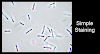
Result Interpretation of Simple Staining
Bacilli and diplobacilli: Rod-shaped bacteria, purple
Spirilla: spiral-shaped bacteria, purple
Cocci: spherical-shaped, bacteria, purple


0 Comments